Lower back pain and hip pain can be debilitating and can significantly impact your quality of life. It is estimated that up to 80% of adults will experience lower back pain at some point in their lives. The causes of lower back pain and hip pain can range from muscle strain, poor posture, and injury to more serious conditions such as arthritis, sciatica, and herniated discs. Fortunately, there are a variety of exercises that can help to reduce pain and improve mobility. In this article, we will discuss the causes of lower back pain and hip pain, as well as exercises that can be used to provide relief.
Understanding the Causes of Lower Back Pain and Hip Pain: What You Need to Know
Lower back pain and hip pain can be caused by a variety of factors, ranging from poor posture to medical conditions. Understanding the causes of lower back pain and hip pain is essential for finding the right treatment and preventing future episodes. In this article, we’ll discuss the most common causes of lower back pain and hip pain, as well as the treatments available.
Poor posture is one of the most common causes of lower back pain and hip pain. Sitting for long periods of time, slouching, and carrying heavy items can all lead to poor posture, which can cause the muscles and ligaments in the lower back and hips to become strained. Additionally, poor posture can lead to misalignment of the spine, which can cause pain in the lower back and hips.
Injury is another common cause of lower back pain and hip pain. Injury to the muscles, ligaments, or tendons in the lower back or hips can cause pain and discomfort. Additionally, injury to the spine, such as a herniated disc, can cause pain in the lower back and hips.
Medical conditions can also cause lower back pain and hip pain. Arthritis, osteoporosis, and sciatica are all conditions that can cause pain in the lower back and hips. Additionally, certain types of cancer can cause pain in the lower back and hips.
The treatment for lower back pain and hip pain depends on the cause. For example, if the pain is caused by poor posture, then the treatment may involve physical therapy, stretching, and strengthening exercises. If the pain is caused by an injury, then the treatment may involve rest, ice, and anti-inflammatory medications. If the pain is caused by a medical condition, then the treatment may involve medications, physical therapy, and surgery.
In conclusion, lower back pain and hip pain can be caused by a variety of factors, ranging from poor posture to medical conditions. Understanding the causes of lower back pain and hip pain is essential for finding the right treatment and preventing future episodes. If you are experiencing lower back pain and hip pain, it is important to speak to your doctor to determine the cause and find the best treatment for your condition.
Exercises to Help Relieve Lower Back Pain and Hip Pain: A Comprehensive Guide
Lower back pain and hip pain can be debilitating and can significantly reduce quality of life. Fortunately, there are a variety of exercises that can help to relieve the pain and improve mobility. This comprehensive guide will provide an overview of the types of exercises that can help to reduce lower back pain and hip pain, as well as tips for how to perform them safely and effectively.
Stretching Exercises
Stretching exercises are an important part of any exercise program for lower back pain and hip pain. Stretching helps to improve flexibility, reduce muscle tension, and improve range of motion.
The following are some examples of stretching exercises that can help to relieve lower back pain and hip pain:
• Knee-to-chest stretch: Lie on your back with your knees bent and your feet flat on the floor. Gently pull one knee up towards your chest, using your hands to hold it in place. Hold the stretch for 30 seconds, then switch legs.
• Hamstring stretch: Lie on your back with your legs straight. Gently pull one leg up towards your chest, using your hands to hold it in place. Hold the stretch for 30 seconds, then switch legs.
• Gluteal stretch: Lie on your back with your knees bent and your feet flat on the floor. Cross one leg over the other, and gently pull the crossed leg towards your chest, using your hands to hold it in place. Hold the stretch for 30 seconds, then switch legs.
• Piriformis stretch: Lie on your back with your knees bent and your feet flat on the floor. Cross one leg over the other, and gently pull the crossed leg towards your chest, using your hands to hold it in place. Hold the stretch for 30 seconds, then switch legs.
• Hip flexor stretch: Kneel on one knee with the other leg bent in front of you. Gently push your hips forward, using your hands to hold them in place. Hold the stretch for 30 seconds, then switch legs.
Strengthening Exercises
Strengthening exercises are also important for reducing lower back pain and hip pain. Strengthening exercises help to improve muscle strength and stability, which can help to reduce pain and improve mobility.
The following are some examples of strengthening exercises that can help to relieve lower back pain and hip pain:
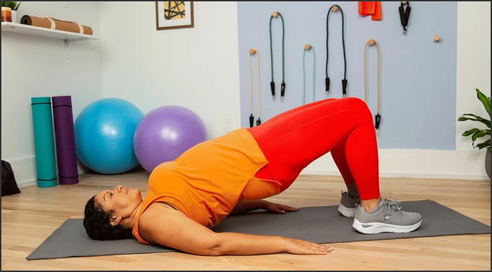
• Bridge: Lie on your back with your knees bent and your feet flat on the floor. Lift your hips off the floor, using your glutes and hamstrings to do the work. Hold the position for 10 seconds, then lower your hips back down.
• Clamshell: Lie on your side with your knees bent and your feet together. Gently lift your top knee up, using your glutes and hip abductors to do the work. Hold the position for 10 seconds, then lower your knee back down.
• Glute bridge: Lie on your back with your knees bent and your feet flat on the floor. Lift your hips off the floor, using your glutes and hamstrings to do the work. Hold the position for 10 seconds, then lower your hips back down.
• Side-lying hip abduction: Lie on your side with your knees bent and your feet together. Gently lift your top leg up, using your glutes and hip abductors to do the work. Hold the position for 10 seconds, then lower your leg back down.
• Wall sit: Stand with your back against a wall and your feet shoulder-width apart. Slowly slide down the wall until your thighs are parallel to the floor. Hold the position for 10 seconds, then slowly slide back up the wall.
Safety Tips
When performing any exercise, it is important to do so safely. Here are some tips for performing exercises safely and effectively:
• Warm up before exercising: Before performing any exercise, it is important to warm up your muscles. This can be done by doing some light stretching or walking for a few minutes.
• Use proper form: Make sure to use proper form when performing any exercise. This will help to ensure that you are getting the most out of the exercise and that you are not putting yourself at risk of injury.
• Listen to your body: If you experience any pain or discomfort while performing an exercise, stop immediately and consult a doctor or physical therapist.
• Progress slowly: When starting a new exercise program, it is important to progress slowly. Start with low-intensity exercises and gradually increase the intensity as your body becomes stronger and more comfortable with the movements.
Conclusion
Lower back pain and hip pain can be debilitating and can significantly reduce quality of life. Fortunately, there are a variety of exercises that can help to relieve the pain and improve mobility. This comprehensive guide has provided an overview of the types of exercises that can help to reduce lower back pain and hip pain, as well as tips for how to perform them safely and effectively. Remember to always warm up before exercising, use proper form, listen to your body, and progress slowly. With the right exercises and proper technique, you can reduce your lower back pain and hip pain and improve your quality of life.Lower back pain and hip pain can be caused by a variety of factors, including poor posture, muscle imbalances, and injury. Fortunately, there are a variety of exercises that can help to reduce pain and improve mobility. These exercises should be tailored to the individual’s specific needs and should be performed regularly for the best results. Additionally, it is important to address any underlying causes of the pain, such as poor posture or muscle imbalances, in order to prevent the pain from returning. With the right exercises and lifestyle modifications, lower back pain and hip pain can be managed and relieved.